Feedback Form

Ultrasound
Diagnostic ultrasound, also called sonography or diagnostic medical sonography, is an imaging method that uses high-frequency sound waves to produce images of structures within your body. The images can provide valuable information for diagnosing and treating a variety of diseases and conditions. Most ultrasound examinations are done using a sonar device outside your body, though some ultrasound examinations involve placing a device inside your body.

Ultrasound is used for a variety of reasons, including to:

  • View the uterus and ovaries of a pregnant woman and assess her fetus
  • Diagnose gallbladder disease
  • Evaluate flow in blood vessels
  • Guide a needle for biopsy or tumor treatment
  • Evaluate a breast lump
  • Check a thyroid gland
  • Diagnose some cancers
  • Reveal genital and prostate abnormalities

Diagnostic ultrasound is a safe procedure that uses low-power sound waves. There are no known risks.

Although ultrasound is a valuable tool, it has limitations. Sound doesn't travel well through air or bone, so ultrasound isn't effective at imaging parts of your body that have gas in them or are hidden by bone. To view these areas, your doctor may order other imaging tests, such as CT or MRI scans or X-rays.

Most ultrasound exams require no preparation, with a few exceptions:

  • For some ultrasound exams, such as of the gallbladder, your doctor may ask that you not eat or drink for up to 6 hours before the exam.
  • Other ultrasound exams, such as of the pelvis, may require a full bladder, so your doctor might ask you to drink up to six glasses of water two hours before the exam and not urinate until the exam is completed.

When scheduling your ultrasound, ask your doctor for specific instructions for your exam.

During an ultrasound exam, you may need to remove jewelry and some or all of your clothing, change into a gown, and lie on an examination table. Gel is applied to your skin to keep air pockets that can block the sound waves from forming.

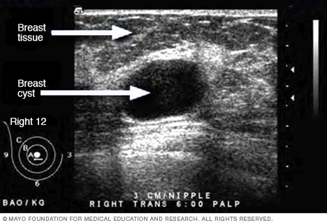
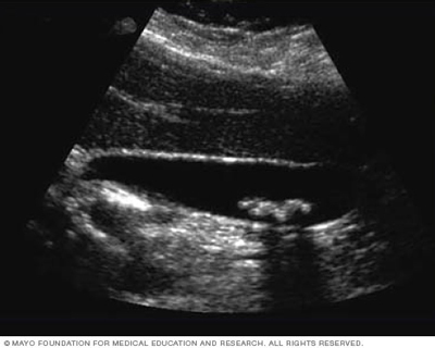
A trained technician (sonographer) presses a small, hand-held device (transducer), about the size of a bar of soap, against your skin over the area being examined, moving it as necessary to capture the image. The transducer sends sound waves into your body, collects sound waves that bounce back and sends them to a computer, which creates the images.

Some ultrasounds are done inside your body. A transducer is attached to a probe that's inserted into a natural opening in your body. Examples of these exams include:

  • Transesophageal echocardiogram. A transducer is inserted into your esophagus, usually with sedation, to obtain heart images.
  • Transrectal ultrasound. A transducer is inserted into a man's rectum to view his prostate.
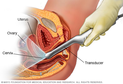
  • Transvaginal ultrasound. A transducer is inserted into a woman's vagina to view her uterus and ovaries.

Ultrasound is usually painless. However, you may experience mild discomfort as the sonographer guides the transducer over your body, especially if you're required to have a full bladder.

A typical ultrasound exam takes from 30 minutes to an hour.


When your exam is complete, a physician trained to interpret imaging studies (radiologist) analyzes the images and sends a report to your doctor. Your doctor will share the results with you.

You should be able to return to normal activities immediately after an ultrasound.