An eye exam usually involves these steps:
-
First, you'll be asked about your medical history and any vision problems you might be experiencing.
-
Next, how clearly you can see (visual acuity) is measured. This helps determine your prescription for glasses.
-
Your eye pressure is measured, for which you may receive drops that enlarge your pupils.
-
Your eye doctor checks the health of your eyes, possibly using several lights to evaluate the front of the eye and inside of each eye.
-
Finally, your eye doctor discusses what he or she found during the exam and answers questions you have about your eyes.
Part of the examination, such as taking your medical history and the initial eye test, may be performed by a technician who assists your doctor.
Several different tests may be performed during the eye exam. The tests are designed to check your vision and to examine the appearance and function of all parts of your eyes.
Eye muscle test
This test examines the muscles that control eye movement, looking for weakness, poor control or poor coordination. Your eye doctor watches your eye movements as you follow a moving object, such as a pen or light, with your eyes.
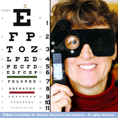
Visual acuity test
This test measures how clearly you see. Your doctor will ask you to identify different letters of the alphabet printed on a chart (Snellen chart) or a screen positioned some distance away. The lines of type get smaller as you move down the chart. Each eye is tested separately. Your near vision also may be tested, using a card with letters similar to the distant eye chart that is held at reading distance.
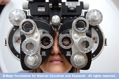
Refraction assessment
Light waves are bent as they pass through your cornea and lens. If light rays don't focus perfectly on the back of your eye, you have "refractive error." Having refractive error may mean you need some form of correction, such as glasses, contact lenses or refractive surgery, to see as clearly as possible. Assessment of your refractive error or refraction helps your doctor determine a lens prescription that will give you the sharpest, most comfortable vision. Refraction assessment may also determine that you don't need corrective lenses.
Your doctor may use a computerized refractor to measure the curve of the surface of your eyes and estimate your prescription for glasses or contact lenses. Or he or she may use a technique called retinoscopy. In this procedure, the doctor shines a light into your eye and measures the refractive error by evaluating the movement of the light reflected by your retina back through your pupil.
Your eye doctor usually fine-tunes this refraction assessment by having you look through a mask-like device that contains wheels of different powers of lenses (phoropter). You'll be asked to judge which combination of lenses gives you the sharpest vision. By repeating this step several times, your doctor finds the lenses that give you the sharpest visual acuity.
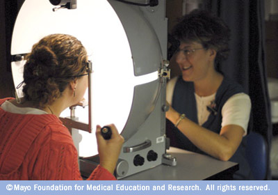
Visual field test (perimetry)
Your visual field is the full extent of what you can see to the sides without moving your eyes. The visual field test determines whether you have difficulty seeing in any areas of your overall field of vision. There are different types of visual field tests:
-
Confrontation visual field exam. Your eye doctor sits directly in front of you and asks you to cover one eye. You look directly at your eye doctor while he or she moves his or her hand in and out of your visual field. You tell your doctor when you can see his or her hand or fingers and how many fingers you see.
-
Tangent screen exam. You sit a short distance from a screen and focus on a target at its center. You tell your doctor when you can see an object move into your peripheral vision and when it disappears.
-
Automated perimetry. Your eye doctor or a technician uses a computer program that flashes small lights as you look into a special instrument. You press a button when you see the lights.
Using your responses to one or more of these tests, your eye doctor determines the fullness of your field of vision. If you aren't able to see in certain areas, noting the pattern of your visual field loss may help your eye doctor diagnose your eye condition.
Color vision testing
You could have poor color vision and not even realize it. If you have difficulty distinguishing certain colors, your eye doctor may screen your vision for a color deficiency. To do this, your doctor shows you several multicolored dot-pattern tests. If you have no color deficiency, you'll be able to pick out numbers and shapes from within the dot patterns. However, if you do have a color deficiency, you'll find it difficult to see certain patterns within the dots. Your doctor may use other tests, as well.
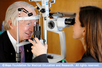
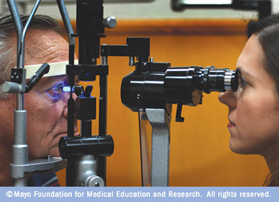
Slit-lamp examination
A slit lamp is a microscope that magnifies and illuminates the front of your eye with an intense line of light. Your doctor uses this light to examine the eyelids, lashes, cornea, iris, lens and fluid chamber between your cornea and iris
When examining your cornea, your doctor may use a dye, most commonly fluorescein (flooh-RES-een), to color the film of tears over your eye and see any damaged cells on the front of your eye. Your tears wash the dye from the surface of your eye fairly quickly.
Retinal examination
A retinal examination — sometimes called ophthalmoscopy or funduscopy — allows your doctor to evaluate the back of your eye, including your retina, optic disk and the underlying layer of blood vessels that nourish the retina (choroid). Usually before your doctor can see these structures, your pupils must be dilated with eyedrops that keep the pupil from getting smaller when your doctor shines light into the eye.
After administering eyedrops and giving them time to work, your eye doctor may use one or more of these techniques to view the back of your eye:
-
Direct examination. Your eye doctor uses an ophthalmoscope to shine a beam of light through your pupil and to see the back of your eye. Sometimes eyedrops aren't necessary to dilate your eyes before this exam.
-
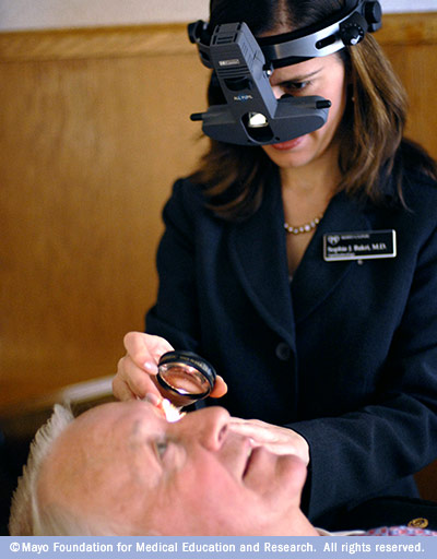
Indirect examination (indirect ophthalmoscopy). During this exam, you might lie down, recline in a chair or sit up. Your eye doctor examines the inside of the eye with the aid of a condensing lens and a bright light mounted on his or her forehead — a bit like a miner's lamp. This exam lets your eye doctor see the retina and other structures inside your eye in great detail and in three dimensions.
-
Slit-lamp exam. In this exam your doctor shines the beam of a slit lamp through a special lens into your eyes. The slit lamp reveals a more-detailed view of the back of your eye.
The retinal examination usually takes less than 10 minutes, but it may take several hours for the effects of the dilating drops to wear off. Your vision will likely be blurry, and you may have trouble focusing on near objects. If light bothers you, you may need to wear dark glasses (or sunglasses) for a short time. You may be uncomfortable driving with dilated pupils, so make sure you have transportation after your exam. Depending on what you need to see at work, you might need to wear reading glasses or remove glasses that correct for nearsightedness until the effects of the eyedrops wear off.
Screening for glaucoma
A test measures your intraocular pressure (tonometry) — the fluid pressure inside your eyes. It helps your eye doctor detect glaucoma, a disease that damages the optic nerve. However, eye pressure measurement is just one thing your eye doctor considers when determining your risk of glaucoma. It's possible to have higher than average eye pressure and not have glaucoma or to have glaucoma if you have normal eye pressure. Your eye doctor will also carefully evaluate your optic nerve for signs of damage.
Methods your eye doctor may use to measure intraocular pressure include:
-
Applanation tonometry. This test measures the amount of force needed to temporarily flatten a part of your cornea. Fluorescein, the same dye used in a regular slit-lamp exam, is usually put in your eye to make your eye easier to see. You'll also receive eyedrops containing an anesthetic. Using the slit lamp, your doctor moves the tonometer to touch your cornea and determine the eye pressure. Because your eye is numbed, you won't feel anything.
-
Noncontact tonometry. This method uses a puff of air to estimate the pressure in your eye. No instruments will touch your eye, so you won't need an anesthetic. You'll feel a momentary pulse of air on your eye, which can be a surprising.
If your eye pressure is higher than average or your optic nerve looks unusual, you doctor may use pachymetry. This test uses sound waves to measure the thickness of your cornea. The most common way of measuring corneal thickness is to put an anesthetic drop in your eye, then place a small probe in contact with the front surface of the eye. The measurement takes seconds.
You may need more-specialized tests, depending on your age, medical history and risk of developing eye disease.